
Giemsa staining - G banding
Before chromosomes can be stained using G banding technique we have to cultivate cells from which we will get chromosomes in condensed state. Sample cells can be any somatic cell (peripheral blood, bone marrow, epithelial cells etc.). Cell culture involves 24 h or 48 h incubation at 37°C in RPMI 1640 medium, enriched with 25% fetal bovine serum and 0.002M L-glutamine. Cultivation of cells sample is done in nutrient medium and then their cell division cycles are stopped and after that chromosomes are stained. Chromosomes are captured in metaphase of mitosis (cell division) by using some cytostatic like colchicine or colcemide.
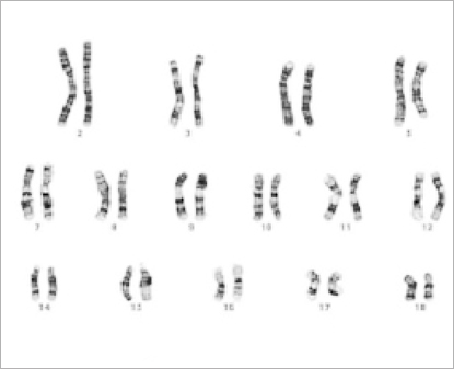
Part of the human karyogram depicting chromosomes with G bands.
G banding or Giemsa banding is a technique used in cytogenetics to produce a visible karyotype (group of all chromosomes) by staining condensed chromosomes. The preparations are stained with a 2% solution of Giemsa in phosphate buffer (pH 6.8) for 3 minutes. It is useful for identifying genetic diseases through the photographic representation of the entire chromosome complement. [8] The metaphase chromosomes are treated with trypsin (to partially digest the histones - proteins that envelope chromosome) and stained with Giemsa stain. Heterochromatic regions, which tend to be rich with adenine and thymine (AT-rich) DNA and relatively gene-poor, stain more darkly in G-banding. In contrast, less condensed chromatin (Euchromatin)—which tends to be rich with guanine and cytosine (GC-rich) and more transcriptionally active—incorporates less Giemsa stain, and these regions appear as light bands in G-banding. The pattern of bands are numbered on each arm of the chromosome from the centromere to the telomere. This numbering system allows any band on the chromosome to be identified and described precisely. [8a]
Cytogenetic analysis and microscope photographing is performed after chromosome preparation and staining according to MIC (Medical Information Center) cooperative group by analyzing 20 cells in metaphase. Analysis can detect structural (for example translocations, deletions, additions etc.) and numeric (change in the number of individual chromosomes like disomy, trisomy etc. and change in number of chromosome garnitures - polyploidy) aberrations of chromosomes.
© www.humankaryotype.com