
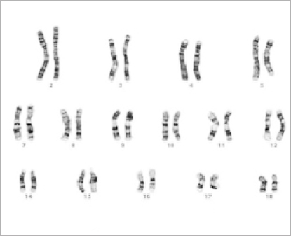
Testing methodology
Until 1920, the methodology commonly used for chromosomal analysis was based on tissue fixation in Carnoy-Flemming solution, preparation of microtomic sections by paraffin process, and staining the preparations with iron hematoxylin. Many of these chromosomal analyzes were performed on testicular tissue taken from patients with tuberculosis of the epididymis or from executed criminals. Therefore, it seems likely that the processing of these tissues by fixation, dehydration, paraffining, and high temperatures as well as posthumous changes, played an important role in modulating the variability of the results of chromosomal analysis. These methods provided insight into "mitotic figures", with a significant overlap of chromosomes in the dense metaphase plate and a low level of differentiation of their morphology. It is therefore not surprising that scientists of the time had different ideas about the number of human chromosomes. In 1955, Tjio and Levan published a paper with the exact number of human chromosomes (2n = 46) based on the culture of embryonic lung cells.
During the second half of the 20th century, in less than 50 years, there have been two revolutions in biology one in protein biochemistry followed by one in molecular genetics with major repercussions both for fundamental research and medical applications. The development of protein biochemistry (at the end of the 1950s) has led to the purification of many proteins followed by the study using crystallography of their three-dimensional structure and, in parallel with this, their sequencing (identification of the linking of amino acids). [6]
The consequences of the second revolution – in molecular genetics – have been more important in the domain of fundamental research than in its applications, simply because it gives direct access to the genes which direct biological phenomenon whereas before it was only possible, at best, to access their products or just the phenotypic perturbations of the phenomena resulting from mutations. Molecular genetics has allowed the cloning of genes, their sequencing and, above all, the ability to isolate them in vitro, in vectors, to modify them in a targeted manner, and to reintroduce them into cells or organisms to study, in a controlled manner, the way in which they control one or other aspect of a phenomenon. Genetics, in becoming molecular, has very rapidly become an integral part of most biological fields, not only due to its power to dissect a biological phenomenon but also because of its increasing ability to unify disciplines which had previously appeared different or unrelated. [6]
For diagnosis, molecular biologists mostly study and manipulate DNA, but RNA can also be studied, for example in virology where the genomes of many viruses are made of RNA. There are several different divisions of the analysis and testing methods of human chromosomes themselves. Several methods that fall into the domain of cytogenetics and molecular genetics will be briefly described on this site. These methods are as follows:

© www.humankaryotype.com